Transforming The Way
Mental Health Is Treated
Our brain-based approach goes beyond traditional psychiatry to give you the answers you want and the results you deserve.
Talk To A Care Coordinator


IMAGINE how your life would
change if you could overcome…
Anxiety
Attention Deficit Disorder (ADD)
Behavioral Addictions
Bipolar Disorder
Complex Conditions
Concussions & Traumatic Brain Injury
Depression
Drugs and Alcohol Addiction
Memory Problems & Dementia
Post-Traumatic Stress Disorder
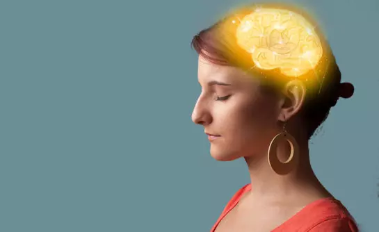
We Take The Guesswork Out of Psychiatry
Comprehensive Evaluation
We take an integrative approach to diagnosis that assesses the biological, psychological, social and spiritual factors that influence your wellbeing.
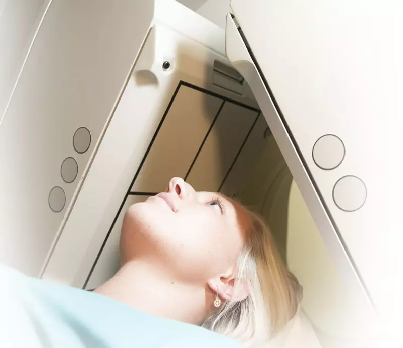
Brain SPECT Imaging
Unlike traditional psychiatry that rarely looks at the organ it treats, we use brain SPECT imaging to help us more accurately diagnose and treat your needs.
Natural Ways to Heal Your Brain
At Amen Clinics, we believe in providing personalized treatment plans that use the least toxic, most effective solutions.
Brain Healthy Life
We provide you with the tools you need to have a better brain and a better life — now and in the future.